Labelled Diagram Of Joints
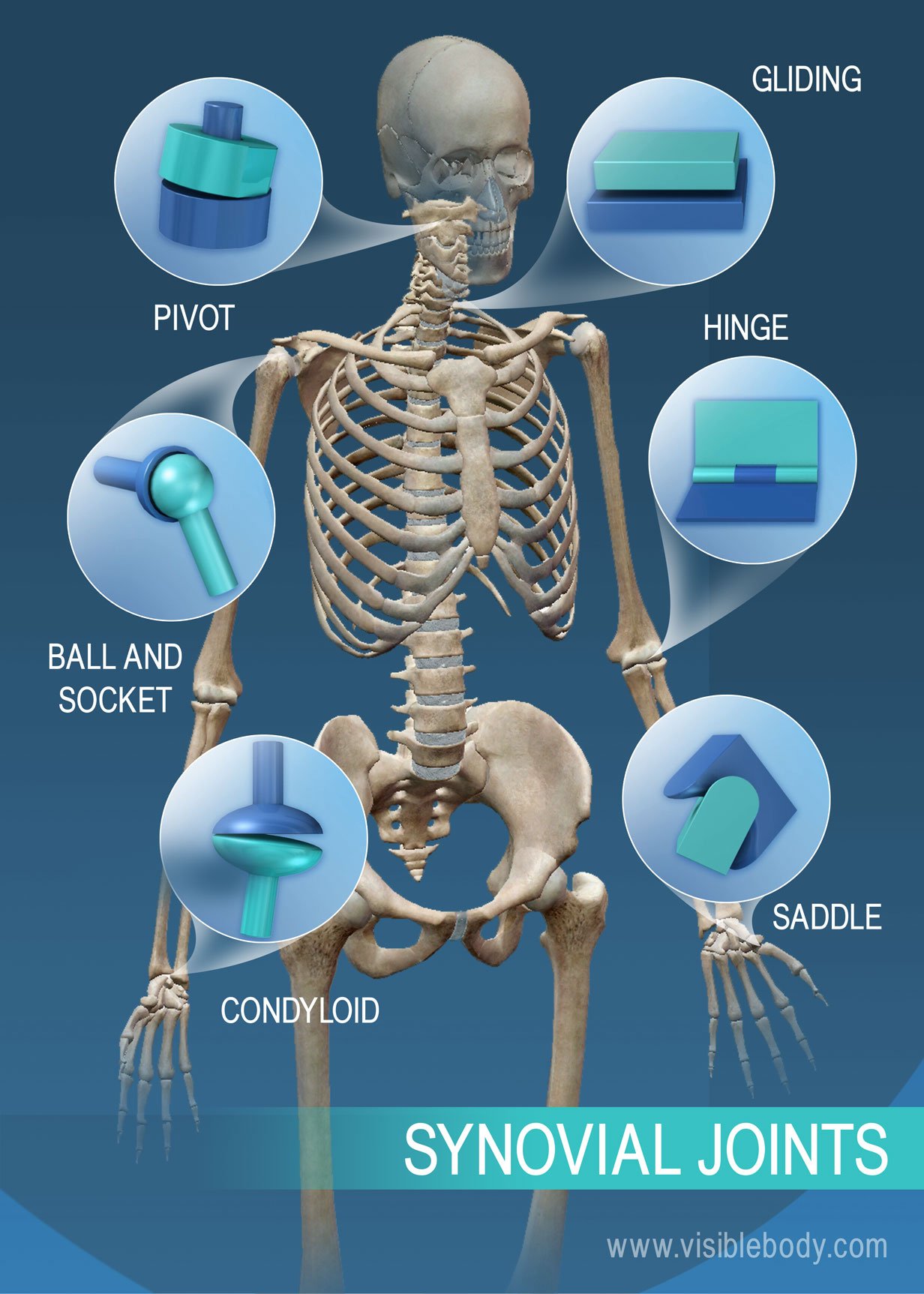
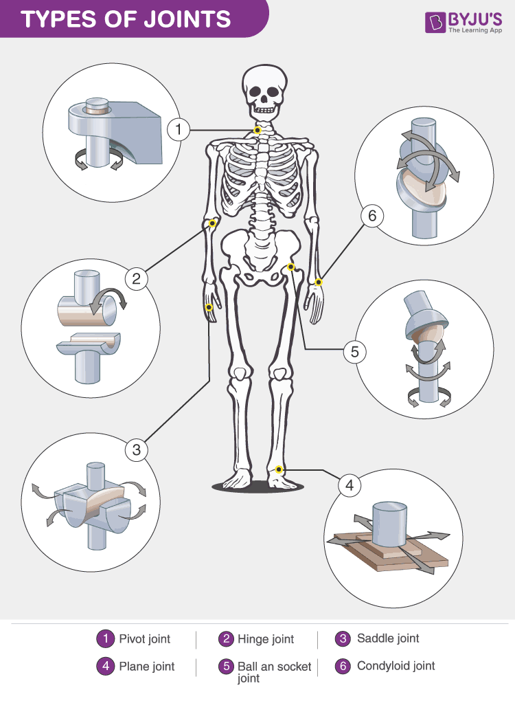
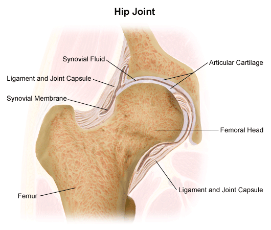
The bones together make up the hip. A hinge joint allows extension and retraction of an appendage.
The articular and epiphyseal branches of the neighboring arteries form a periarticular arterial plexus.
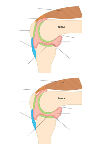
Labelled diagram of joints. Hinge joints are found in the knees elbows fingers and toes. Joints in the human skeleton can be grouped by function range of motion and by structure material. Lubricated with synovial fluid the cartilage forms slippery surfaces for free movements.
Ball and socket joints are found in the shoulders and hips. This articular cartilage is avascular non nervous and elastic. The structures necessary to create this joint are the socket the joint capsule muscle ligaments and the necktrochanter of the femur.
Synovial joints are most evolved and therefore most mobile type of joints. The basic structure of a synovial joint is shown in the diagram below. The main parts of synovial joints are labelled on the synovial joint diagram.
The column runs from the cranium to the apex of the coccyx on the posterior aspect of the body. Synovial joints allow for smooth movements between the adjacent bones. A synovial joint or diarthrosis occurs at articulating bones to allow movement.
That is an organization of joints by structure. The second way to categorize joints is by the material that holds the bones of the joints together. The blood supply of a synovial joint comes from the arteries sharing in anastomosis around the joint.
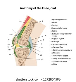
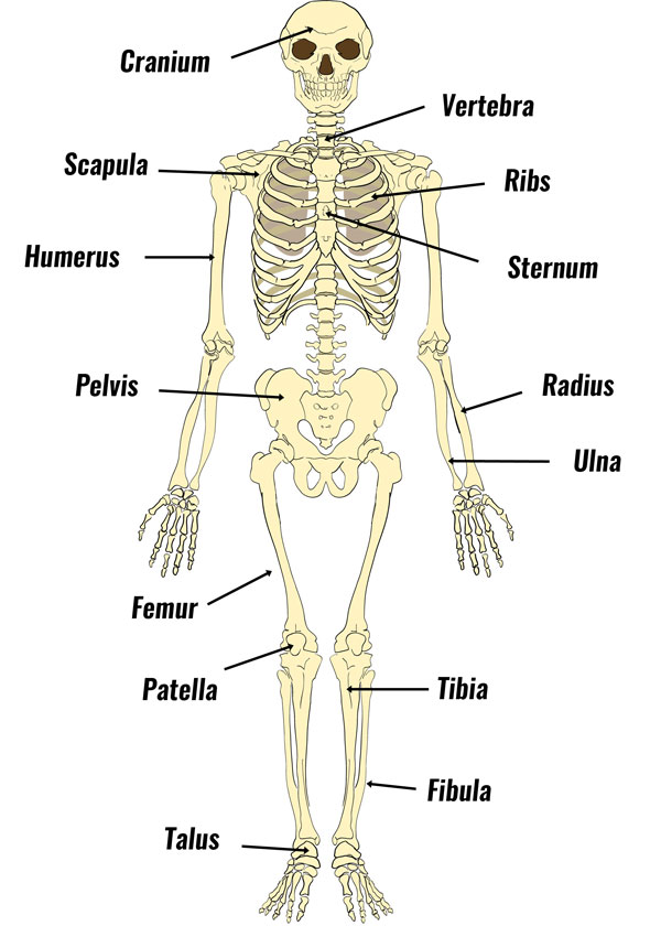
The skeleton acts as a scaffold by providing support and protection for the soft tissues that make up the rest of the body. The pubis ischium and ilium together constitute the pelvis while the thigh bone is the femur. This diagram shows the location of the bursae which are fluid filled sacs in a bone.
It contains and protects the spinal cord. The skeletal system includes all of the bones and joints in the body. The hip itself is a ball and socket joint much like the shoulder.
Its complexity and its efficiency is the best example of gods creation. The articular capsule is highly innervated but avascular lacking blood and. They possess the following characteristic features.
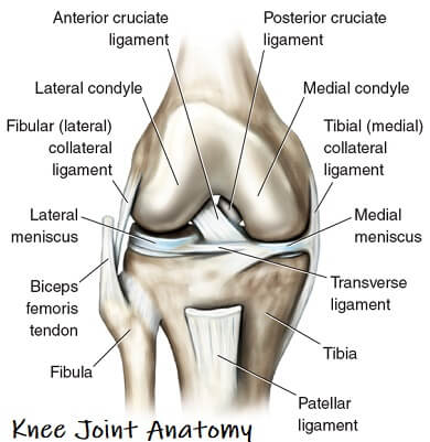
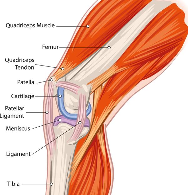
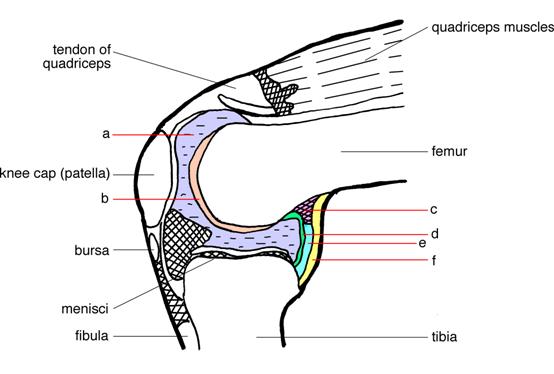
Labeled diagram of the knee joint knee joint is one of the most important hinge joints of our body. Fibrous connective tissue found in various parts of the body such as the joints. The vertebral column also known as the backbone or the spine is a column of approximately 33 small bones called vertebrae.
A ball and socket joint allows for radial movement in almost any direction. The basic structure of a synovial joint is shown in the diagram below. The main parts of synovial joints are labelled on the synovial joint diagram.
There articular surfaces are covered with hyaline cartilage. Each bone is a complex living organ that is made up of many cells protein fibers and minerals. Diagram of the anastomosis around the elbow joint.
The first is by joint function also referred to as range of motion. A saddle joint allows movement back and fourth and up and down.
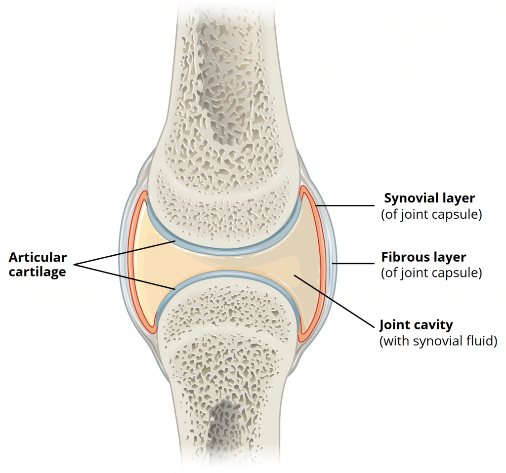 Structures Of A Synovial Joint Capsule Ligaments Teachmeanatomy
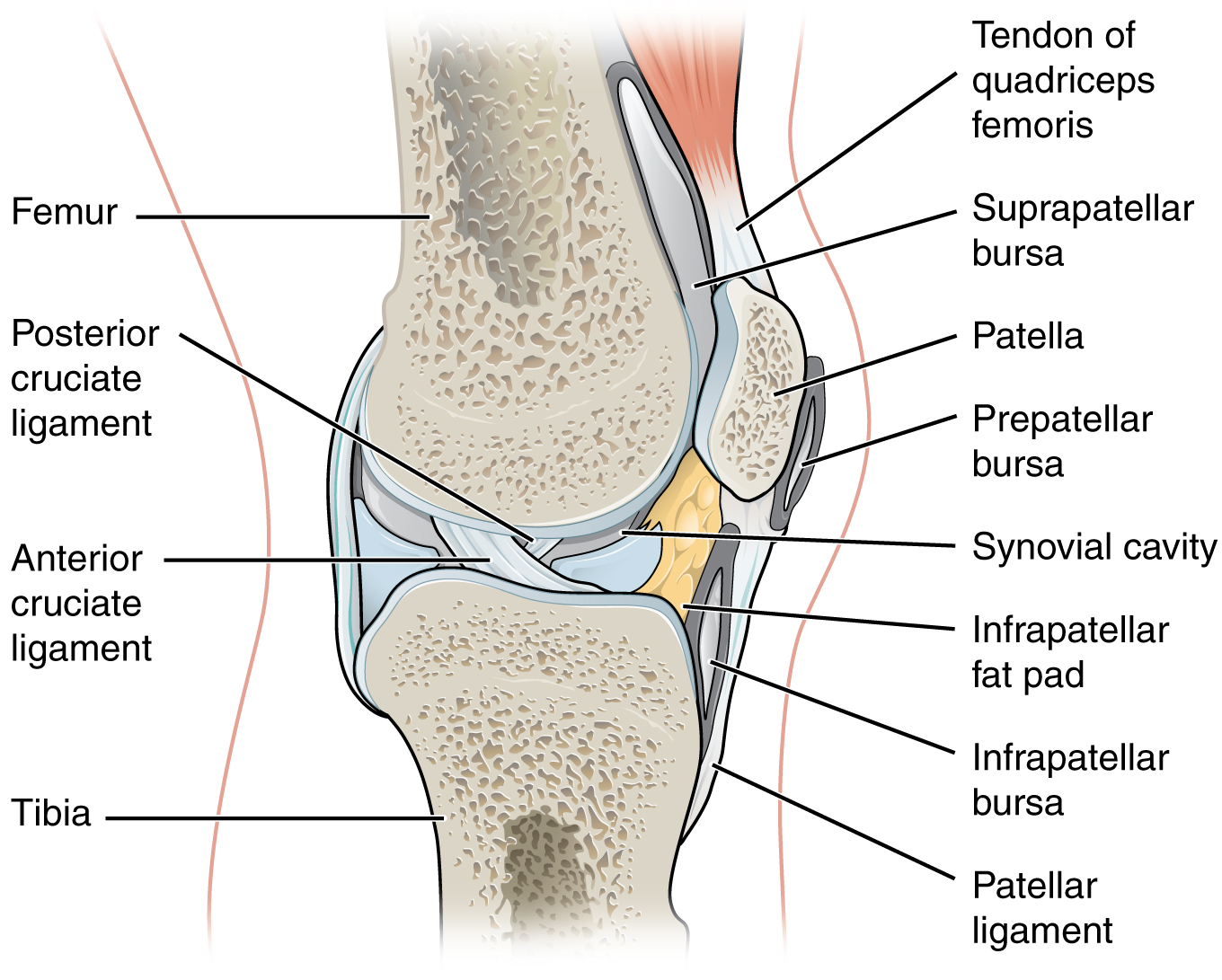 9 4 Synovial Joints Anatomy And Physiology
 9 4 Synovial Joints Anatomy Physiology
 Synovial Joint Structure And Label Diagram Quizlet
 Sketch And Label Typical Synovial Joint Brainly In
 Joint Definition Anatomy Movement Types Britannica
 Knee Joint Labeled Diagram Stock Vector Illustration Of Health
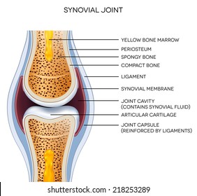 1000 Joint Label Stock Images Photos Vectors Shutterstock
 Structure And Function Of Synovial Joints Hsc Pdhpe
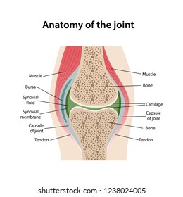 1000 Synovial Joints Stock Images Photos Vectors Shutterstock
 Sketch And Label Typical Synovial Joint Brainly In
 Bones Joints Labelled Diagram Of A Human Elbow Human Health
 Structure Of Synovial Joint Youtube
 A Diagrammatic Explanation Of The Parts Of The Human Knee Bodytomy
Bones Joints And Muscles Archives Medical Information Illustrated
 Labelled Diagram Of Synovial Joint
 Labelled Diagram Of Synovial Joint
 Labelled Diagram Of Bones And Joints Of Hand Bone And Joint
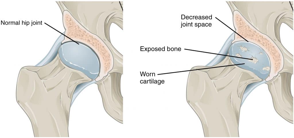 Structures Of A Synovial Joint Capsule Ligaments Teachmeanatomy
Label A Diagram Of The Human Elbow Joint Ib Biology Syllabus
 Synovial Joint Diagram Labeled Stock Vector Illustration Of
 File Human Arm Bones Diagram Svg Wikipedia
Bones Joints And Muscles Archives Medical Information Illustrated
 Anatomy Of Knee Joint Labeled Knee Joint Anatomy Labeled Diagram
 Pivot Joint Definition Examples Function Facts Britannica
 Skeletal System Skeleton Bones Joints Cartilage Ligaments Bursae
11 2 Muscles And Movement Bioninja
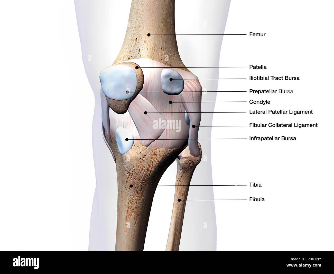 Knee Diagram To Label Unlimited Wiring Diagram
 Skeletal System Skeleton Bones Joints Cartilage Ligaments Bursae
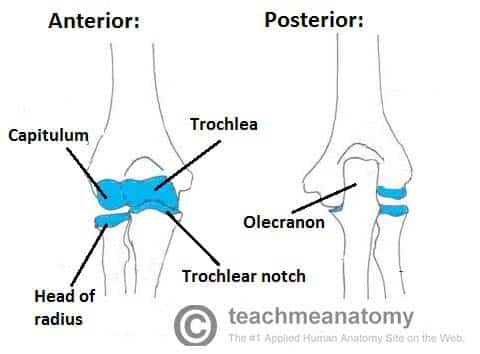 The Elbow Joint Structure Movement Teachmeanatomy
 191 Synovial Joint Stock Illustrations Cliparts And Royalty Free
 Labelled Diagram Of Synovial Joint
 Knee Joint Anatomy Motion Knee Pain Explained
 Knee Joint Cross Section Medical Art Library
 Knee Joint Picture Image On Medicinenet Com
I What Is A Joint Ii Name Four Types Of Joints In The Human Body
 1000 Synovial Joints Stock Images Photos Vectors Shutterstock
19 3 Joints And Skeletal Movement Concepts Of Biology 1st
:background_color(FFFFFF):format(jpeg)/images/library/12804/bones-skeletal-system_english.jpg) Musculoskeletal System Anatomy And Diagram Kenhub
Musculoskeletal System Anatomy And Diagram Kenhub

Anatomy And Physiology Of Animals The Skeleton Wikibooks Open
 Eps Vector Anatomy Structure Knee Joint Vector Stock Clipart
 Anatomy Of The Knee Joint Labelled Royalty Free Cliparts Vectors
 Labelled Diagram Of Synovial Joint
Https Www Msfta Org Cms Lib6 Fl02001163 Centricity Domain 122 Joints Movement Powerpoint Pdf
/shoulder-bones-and-muscles-971624580-9ac67b210b194ca6b414ffc28c8d3402.jpg) Anatomy Of The Human Shoulder Joint
Anatomy Of The Human Shoulder Joint
 A Labeled Diagram Of The Knee With An Insight Into Its Working
 11 1 Essential Ideas 11 1 2 Movement
 Ball And Socket Joint Anatomy Britannica
 Types Of Synovial Joints Learner Support
 Joints And Ligaments Learn Skeleton Anatomy
 Hip Joint Radiology Reference Article Radiopaedia Org
 Skeletal System Labeled Diagrams Of The Human Skeleton
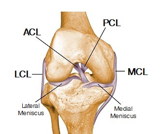 Knee Joint Anatomy Motion Knee Pain Explained
 General Anatomy Of The Bull And The Cow Illustrated Atlas
 Types Of Joints Joint Structures In The Body Video Lesson
 Skeletal System Labeled Diagrams Human Skeleton The Skeletal
 Classification Of Joints Boundless Anatomy And Physiology
:background_color(FFFFFF):format(jpeg)/images/library/12803/musculoskeletal-system.png) Musculoskeletal System Anatomy And Diagram Kenhub
Musculoskeletal System Anatomy And Diagram Kenhub
 Foot Anatomy Detail Picture Image On Medicinenet Com
 Elbow Joint Anatomy Pictures And Information
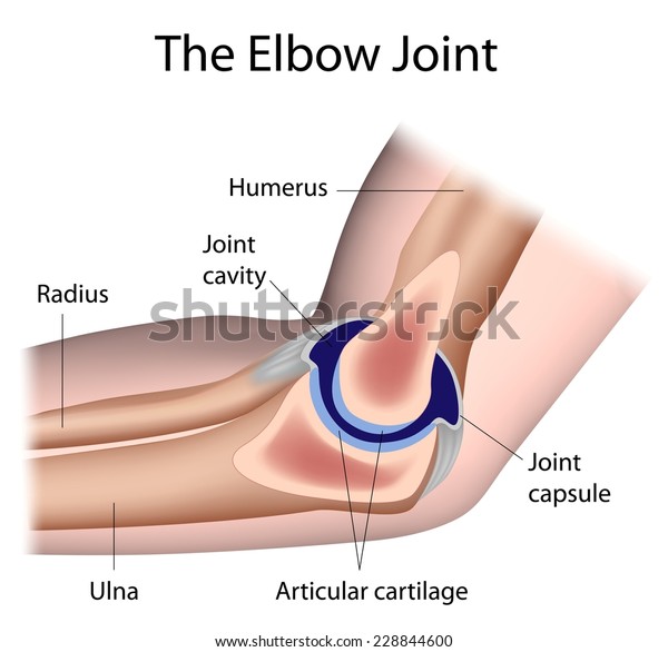 Elbow Joint Anatomy Labeled Science Healthcare Medical Stock Image
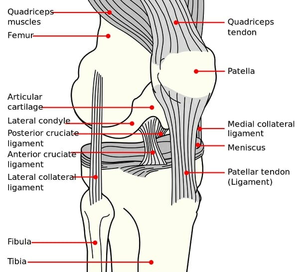 Synovial Joint Diarthrosis Definition Types Structure Examples
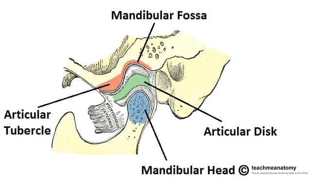 The Temporomandibular Joint Structure Function Teachmeanatomy
 Types Of Joints Classification Of Joints In The Human Body
 The Human Skeleton Bones Structure Function Teachpe Com

 Joints Of Upper Limb Elbow Only Flashcards Quizlet
 Solved 3 The Joints Of A Truss Experience Forces Due To
 Joints Definition Types And Classification Of Joints Videos
Imaging And Quantification Of Joint Shape A Zebrafish 5th Dpf
 Structure And Function Of Synovial Joints Hsc Pdhpe
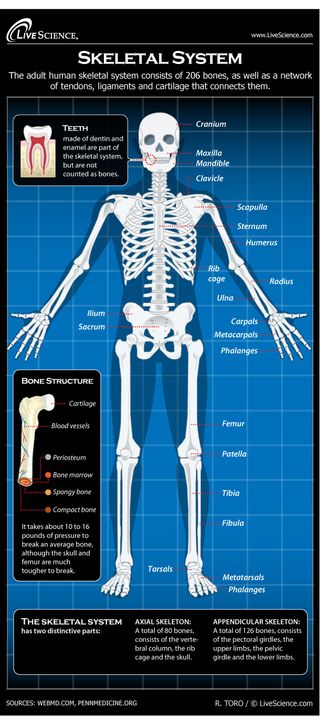 Diagram Of The Human Skeletal System Infographic Live Science
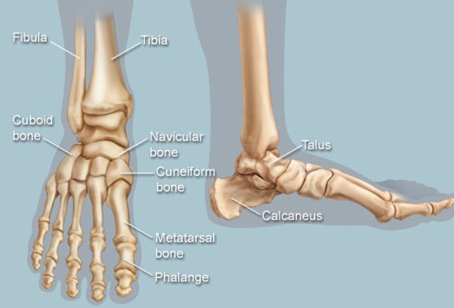 Feet Human Anatomy Bones Tendons Ligaments And More
 Labelled Diagram Of Synovial Joint
 Anatomy Of The Spine Teachpe Com
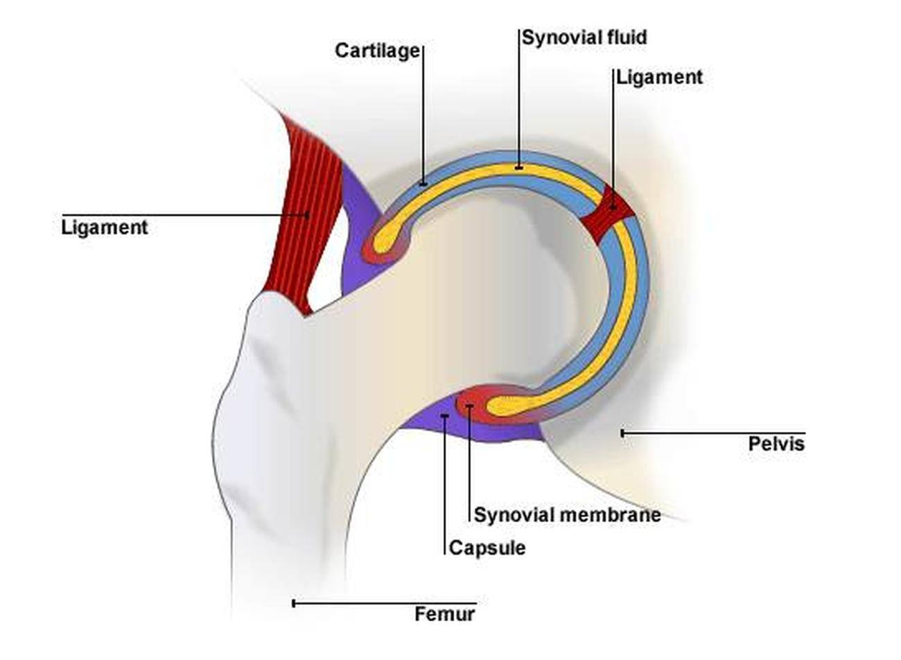 Pictures Of Ball And Socket Joint
Http Www Lamission Edu Lifesciences Alianat1 Chap 206 20 20joints Pdf
 Pelvis Definition Anatomy Diagram Facts Britannica
 General Anatomy Of The Bull And The Cow Illustrated Atlas
Skeletal System Of Human Beings With Diagram
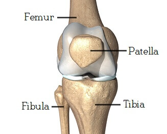 Knee Joint Anatomy Motion Knee Pain Explained
Amphiarthrodial Joints Ground Up Strength
Https Www Pearsonschoolsandfecolleges Co Uk Feandvocational Sportsstudies Alevel Ocralevelpe2008 Samples Samplepagesfromocraspestudentbook Chapter1 Sample Pdf
 Skeletal System Definition Function And Parts Biology Dictionary
 Ligaments Tendons And Joints Video Khan Academy
 Hip Joint Anatomy Pictures And Information
 The Diagram Below Represents One Of The Joints In The Mammalian
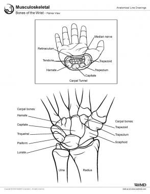 Wrist Joint Anatomy Overview Gross Anatomy Natural Variants
 9 2 Fibrous Joints Anatomy And Physiology
Draw A Neat Diagram Of Ball And Socket Joint Jgv662wjj Science



Komentar
Posting Komentar