Labeled Lysosome Structure Diagram
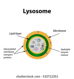
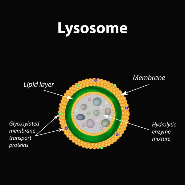
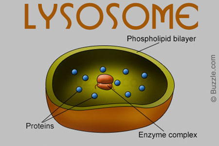
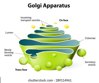
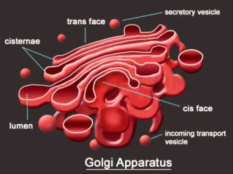
Structure of lysosomes the outer surface is formed by a single membrane a phospholipid bilayer that can fuse with some other membrane bound organelles. Primary lysosomes or proto lysosomes are newly produced organelles bounded by a single membrane and believed to be derived from the trans face of the golgi apparatus.
 Lysosome Images Stock Photos Vectors Shutterstock
The word lysosome was derived from two greek words lysis which means destruction or dissolution and soma which means body.
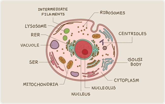
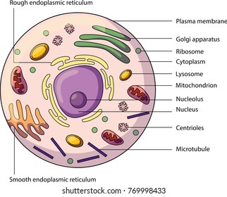
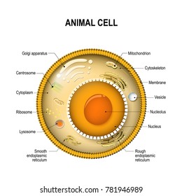
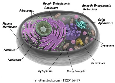
Labeled lysosome structure diagram. Lysosomes are actually membranous sacs filled with enzymes. It is an important cell organelle responsible for inter and extracellular breakdown of substances. Plasma cell membrane cytoplasm mitochondria.
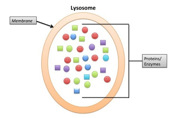
Lysosomes act as garbage disposal of the cell. These are bounded by a single lipoprotein membrane containing enzymes in crystalline form. Lysosomal enzymes are present in the membrane which surrounds the lysosomes.
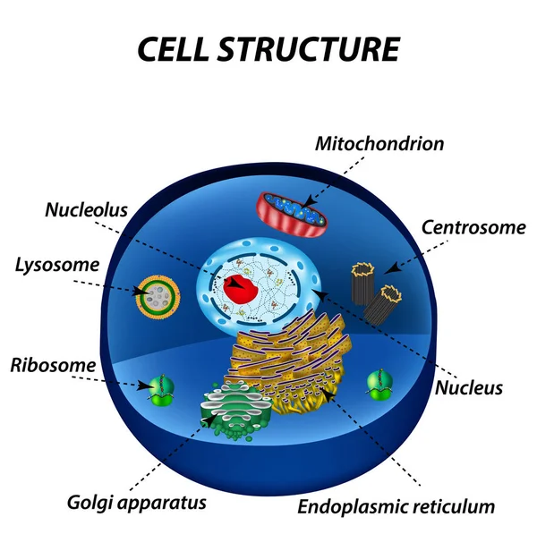
Fflitoehondriae lysosome nucleus endoplasmic reticulumn. 632 lysosome stock photos vectors and illustrations are available royalty free. Lysosomes are membrane bound dense granular structures containing hydrolytic enzymes responsible mainly for intracellular and extracellular digestion.
Lysosomes are found in all eukaryotic cells. Cell structure s function worksheet label and show the locations of the following organelles on the diagram of a plant cell belw. Spherical shape of diameter ranging up to one micrometre 1 mm.
Help us improve your search experience. The word lysosome is made up of two words lysis meaning breakdown and soma meaning body. Plasma membrane forms vesicle around extracellular droplets vesicle formed pinches off into cytosol fusion of lysosome and vesicle digestion by enzymes solutes released.
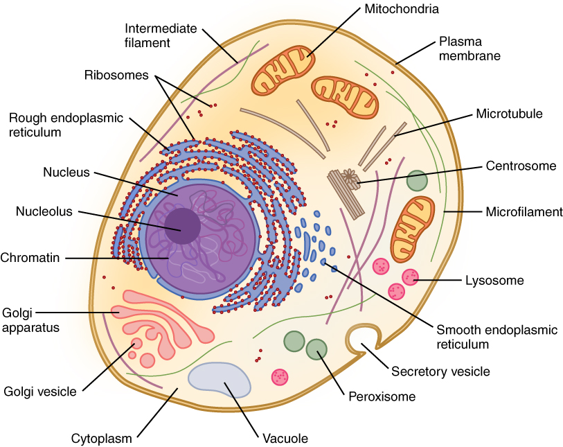
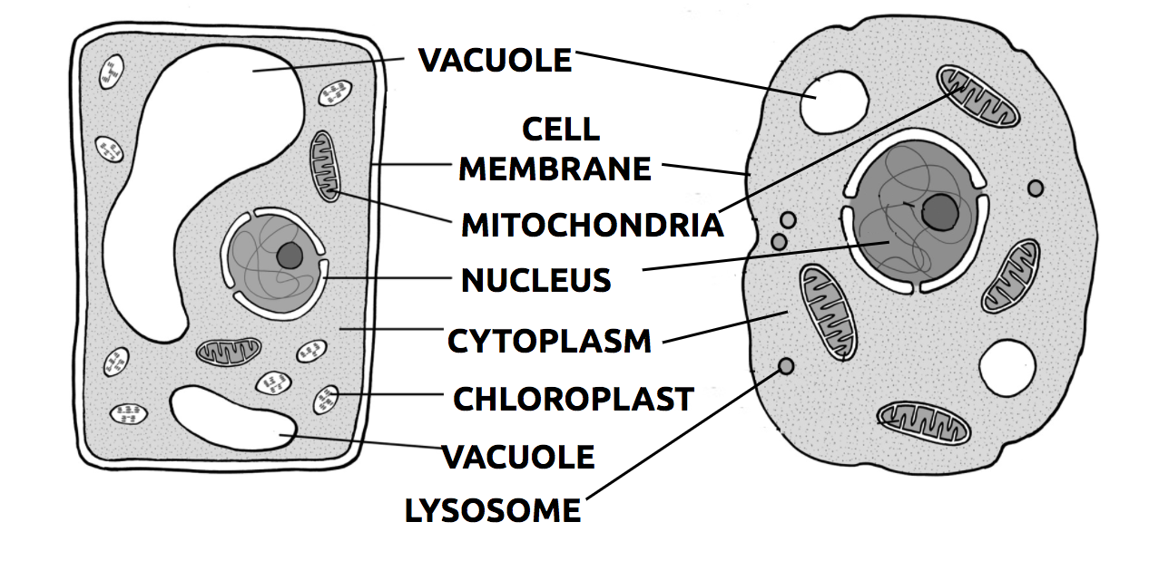
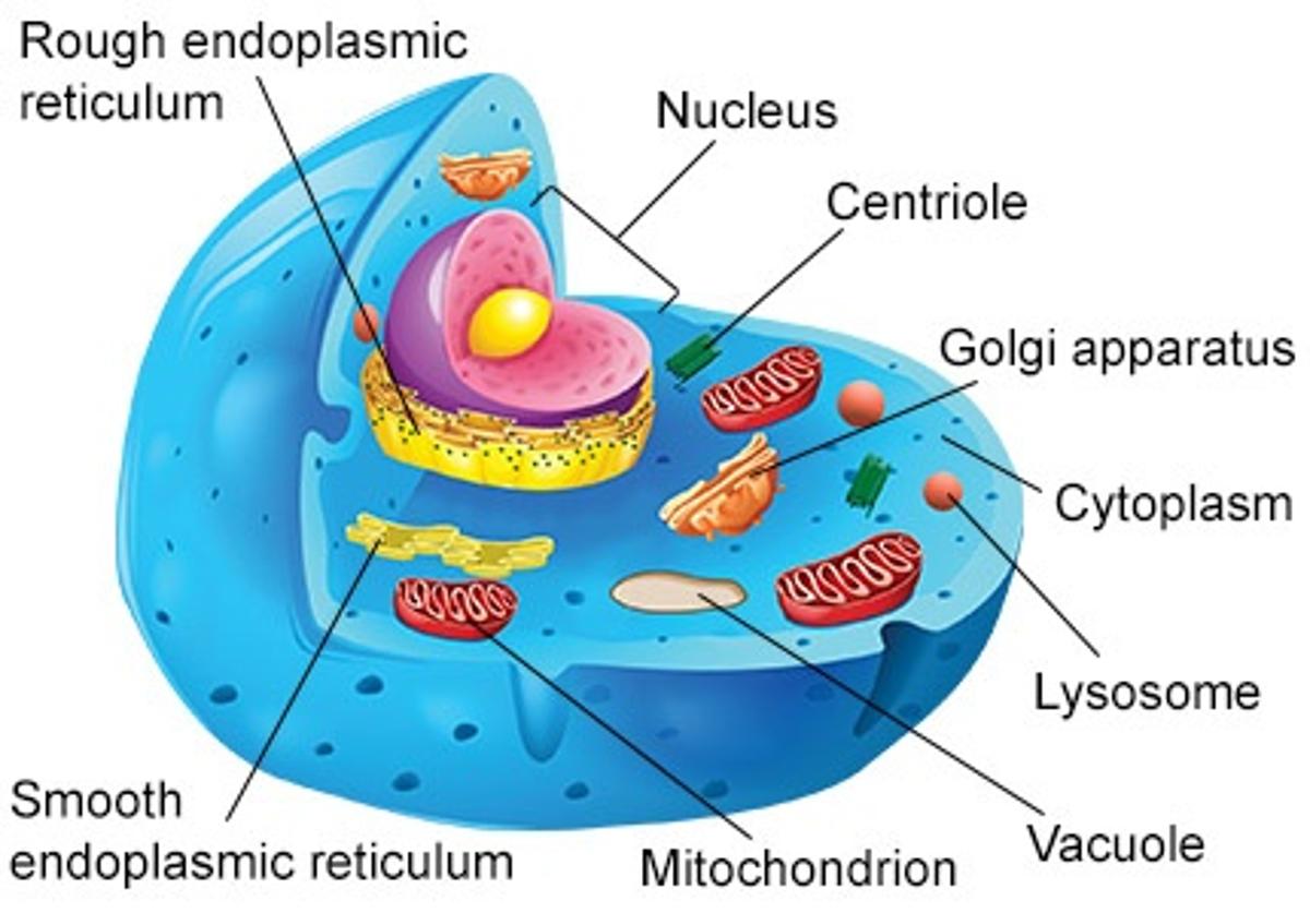
Label and show the locations of the following organelles on the diagram of a animal cell below. Lysosome structure lysosomes are membrane bound organelles and the area within the membrane is called the lumen which contains the hydrolytic enzymes and other cellular debris. Food enzyme animal cell diagram fungus cell diagram of an animal cell lysossomal cell vacuole plant animal cells vacuoles lymosomes structure of a fungus.
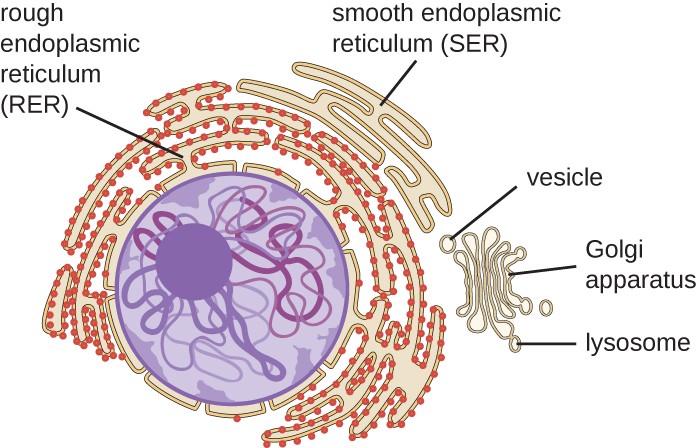
The diagram below shows the lysosome structure within a cell. Lysosomes are sphere shaped flattened structures. Meanwhile the shape and size of lysosomes may differ in different organisms.
The enzymes present are phosphatase nuclease lipase protease glycosidase sulfates amylase. Lysosomes are globular or granular in appearance of 02 05um size without any characteristic shape or structure. The lysosomal structure size ranges from 01 to 12mm.
Lysosomes structure enzymes types functions. Lysosomes were discovered in 1949 by a belgian cytologist christian de duve. Although varying somewhat in size primary lysosomes are typically about 100 nm in diameter.
 Lysosome Images Stock Photos Vectors Shutterstock
Lysosomes Meaning Structure And Function With Diagram
 Lysosome Images Stock Photos Vectors Shutterstock
 Lysosome Images Stock Photos Vectors Shutterstock
 Lysosome Stock Vectors Royalty Free Lysosome Illustrations
 Lysosome Definition Function Structure Biology Dictionary
 Lysosome Description Formation Function Britannica
Animal Cell Model Diagram Project Parts Structure Labeled Coloring
 Lysosome Images Stock Photos Vectors Shutterstock
 Lysosome New World Encyclopedia
 Lysosome Structure Biology Wise
 Image Of A Plant Cell Diagram With Each Organelle Labeled Plant
 The Fascinating World Of Cell Organelles And Their Functions
 Image Of An Animal Cell Diagram With Each Organelle Labeled
 The Nucleus And Cytoplasm Anatomy And Physiology
 Lysosomes Structure Definition Function Diagram
 Lysosome Images Stock Photos Vectors Shutterstock
 Lysosome Structure Biology Wise
 ᐈ Animal Cells Labeled Stock Pictures Royalty Free Organelle

Animal Cell Model Diagram Project Parts Structure Labeled Coloring
Useful Notes On The Four Kinds Of Lysosomes Biology
 What Is A Cell Animal Cell Project Human Cell Structure 3d
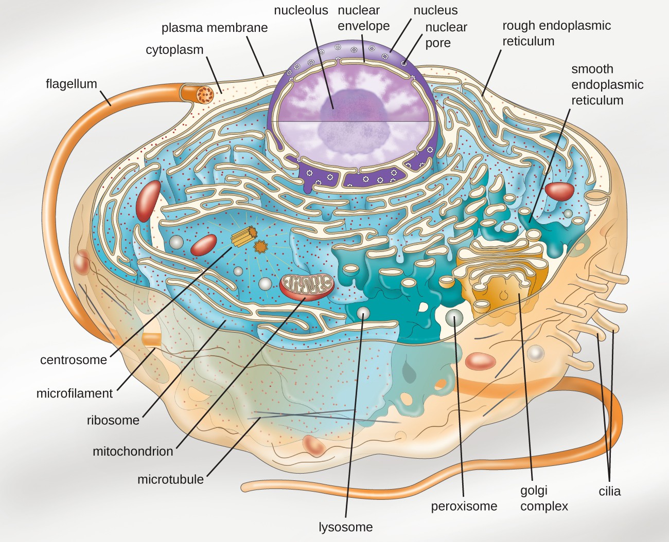 Unique Characteristics Of Eukaryotic Cells Microbiology
 Labeled Animal Cell Physiology
 Lysosome Structure Biology Wise
Animal Cell Structure Diagram Model Animal Cell Parts And
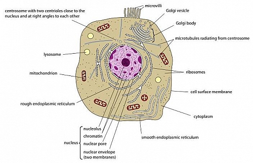 Animal Cell Structure Diagram Model Animal Cell Parts And
 A Labeled Diagram Of The Animal Cell And Its Organelles Biology Wise
 Golgi Apparatus Function Structure With Analogy Diagram
 Review The Diagram Of The Animal Cell Given Below And Answer The
 Eukaryotic Cell Structure Labeling Animal Diagram Quizlet
 Edtb Ko Disrupts Lysosomes Immunofluorescent Labeling Of Ileum
 Eps Vector Amoeba Labeled Vector Illustration Single Cell
 Apex2 Technology For Em Shows Ectopic Expression Of Lamp1 Is
 Animal Cell Diagram Labeled Cakepins Com Animal Cells Model
 Fat Cell Structure Vector Illustration Labeled Anatomical
 Science Source Liver Cell With Labeled Structures Illustration
 Lysosomes Types Morphology Function Process And Microscopy
 Cell Wall Description Properties Components Communication
Cell Structure And Functions With Diagram
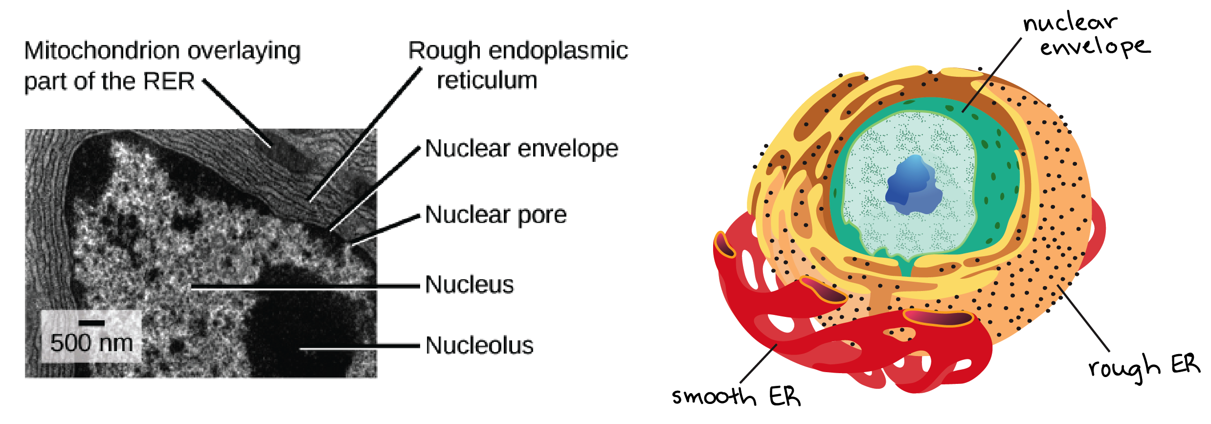 The Endomembrane System Article Khan Academy
 Eukaryotic Cell Structure Sciencetopia
 A Labeled Diagram Of An Animal Cell For Biology Class Animal
Lysosomes And Peroxisomes Celebrate Cytochemistry Gwen V
Main Functions Of The Golgi Apparatus With Diagram
 Immunogold Labeling For The Lysosomal Membrane Protein Lgpl20 A
 Identify The Nine Cell Structures That Are Labeled In The Animal
 Organelle Structure And Function A Level Notes
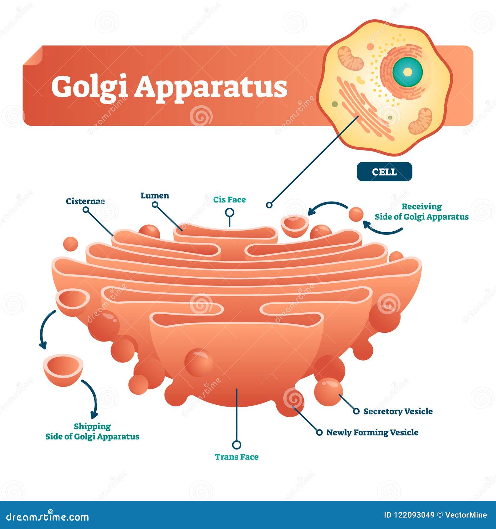 Golgi Apparatus Vector Illustration Labeled Microscopic Scheme
 Eukaryotic Cell Structures Review Article Khan Academy
Plant Cell Anatomy Enchanted Learning
 Animal Cell Images Stock Photos Vectors Shutterstock
Cells Mr Scott S 6th Grade Class
 Quia Ap Chapter 6 Cells Detailed
 Mrl Lpr Mfs Phagocytose And Traffic Igg Ics To Lysosomal
 Human Cell Diagram Human Cell Diagram Cell Diagram
 Golgi Apparatus Definition Function And Structure Biology
 2 3 2 Annotate The Diagram Of A Eukaryotic Cell With The Functions
 Golgi Apparatus Definition Function Location Facts Britannica
 Garp Complex Mutations Lead To Enlarged Lysosomes A Confocal
:max_bytes(150000):strip_icc()/endocytosis-5ad64d57c0647100386364bb.jpg) A Definition Of Endocytosis With Steps And Types
A Definition Of Endocytosis With Steps And Types
 Animal Cell Model Diagram Project Parts Structure Labeled
 Printable Animal Cell Diagram Labeled Unlabeled And Blank
 An In Depth Look At The Structure And Function Of Cytoplasm
 Animal Cell Diagram With Labels By Russell Kightley Media
 Unique Characteristics Of Eukaryotic Cells Microbiology
 Consider This Animal Cell The Organelles In An Animal Cell Are
 Neuronal Soma Derived Degradative Lysosomes Are Continuously
 Animal Cell For Kids Label The Parts Animal Cell Project
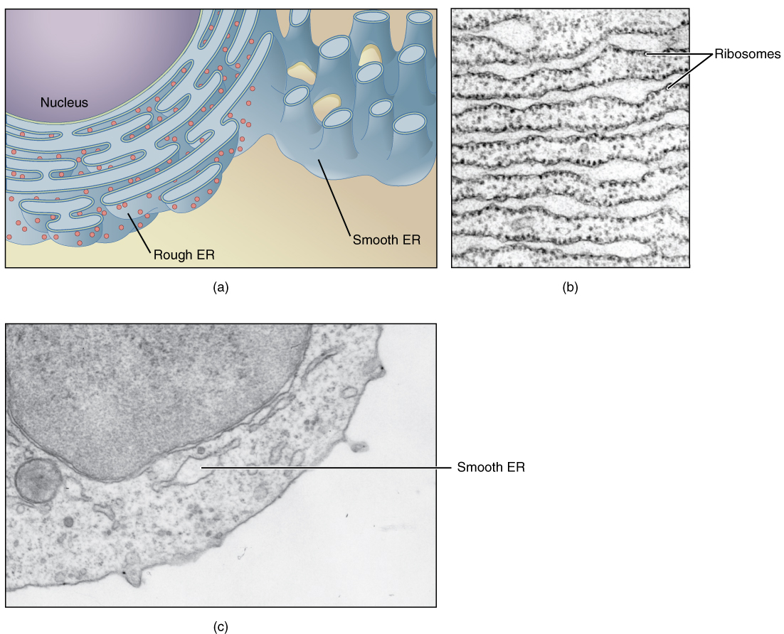 3 2 The Cytoplasm And Cellular Organelles Anatomy And Physiology
:max_bytes(150000):strip_icc()/animal_cell_organelles-36b9ba0c39a44a429ccbb0702ff43d79.jpg) Animal Cells And The Membrane Bound Nucleus
Animal Cells And The Membrane Bound Nucleus
 Amoeba Labeled Vector Illustration Single Cell Animal Structure
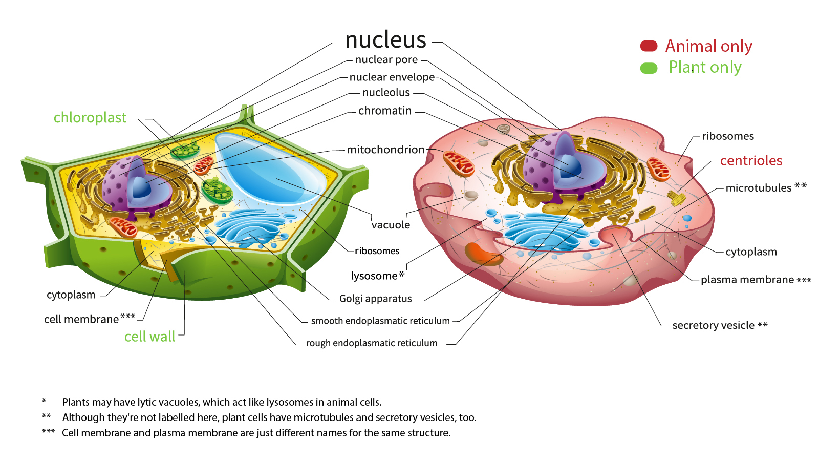 Here S How Plant And Animal Cells Are Different Howstuffworks
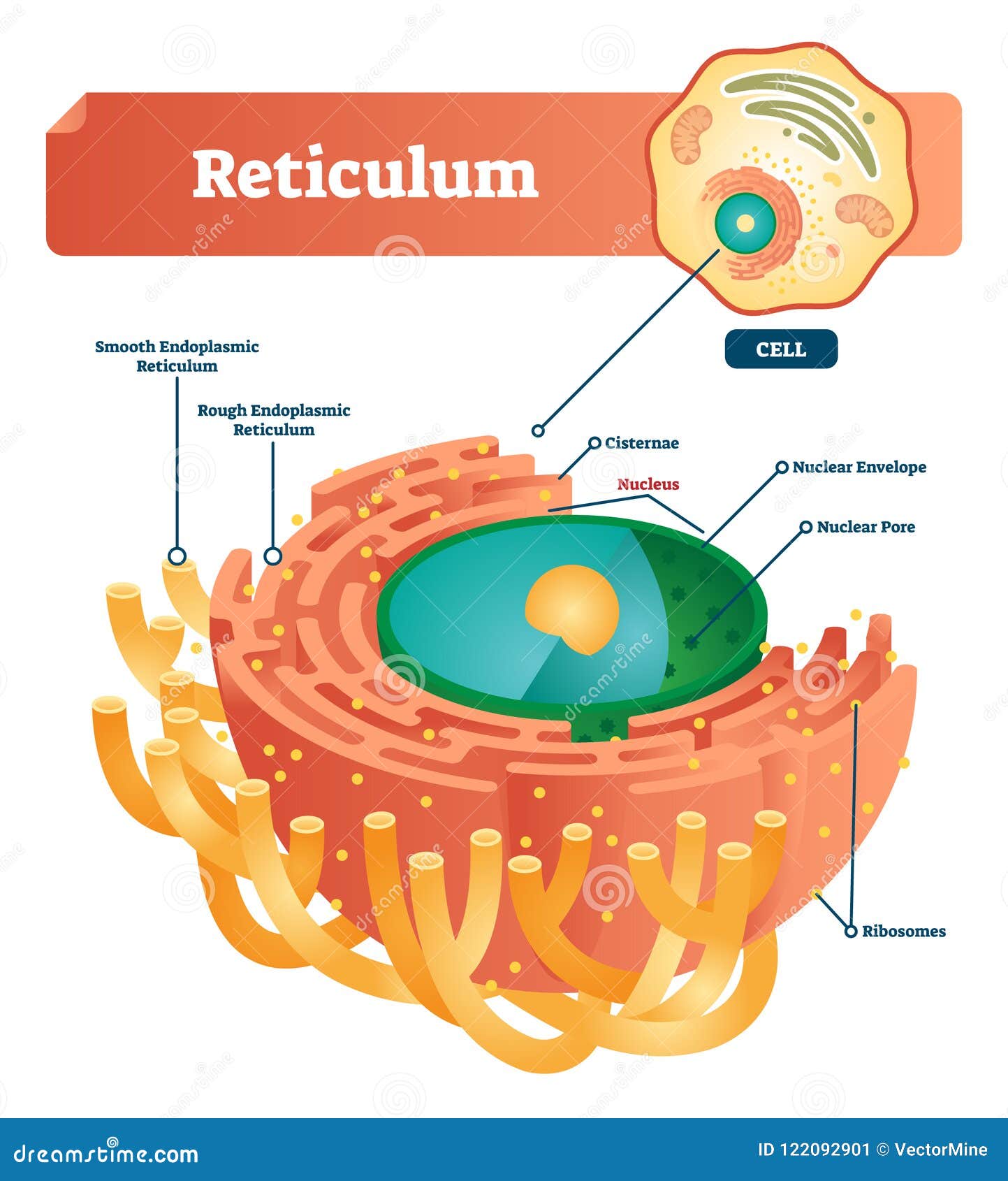 Reticulum Labeled Vector Illustration Scheme Anatomical Diagram
 Labeled Plant Cell With Diagrams Science Trends
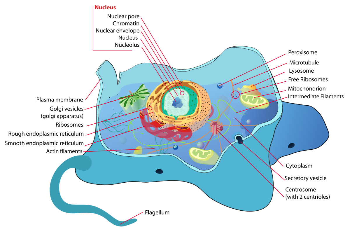 File Animal Cell Structure En Svg Wikimedia Commons
 Eukaryotic And Prokaryotic Cells Similarities And Differences
Animal Cell Model Diagram Project Parts Structure Labeled Coloring
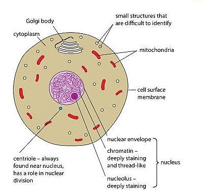 Animal Cell Structure Diagram Model Animal Cell Parts And
 Autophagosome Labeling Lentivirus Map1lc3b Cfp 500 00
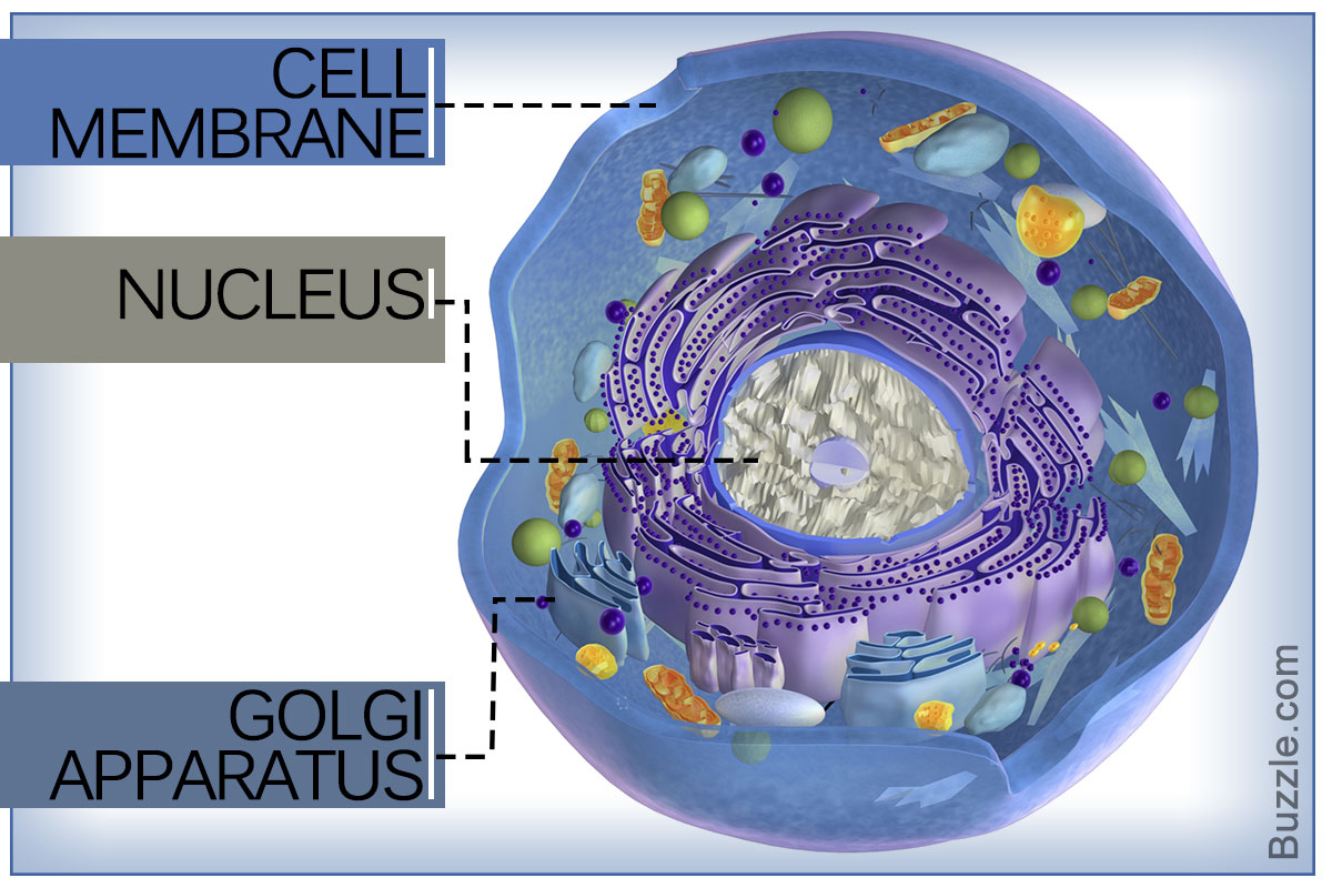 A Labeled Diagram Of The Animal Cell And Its Organelles Biology Wise
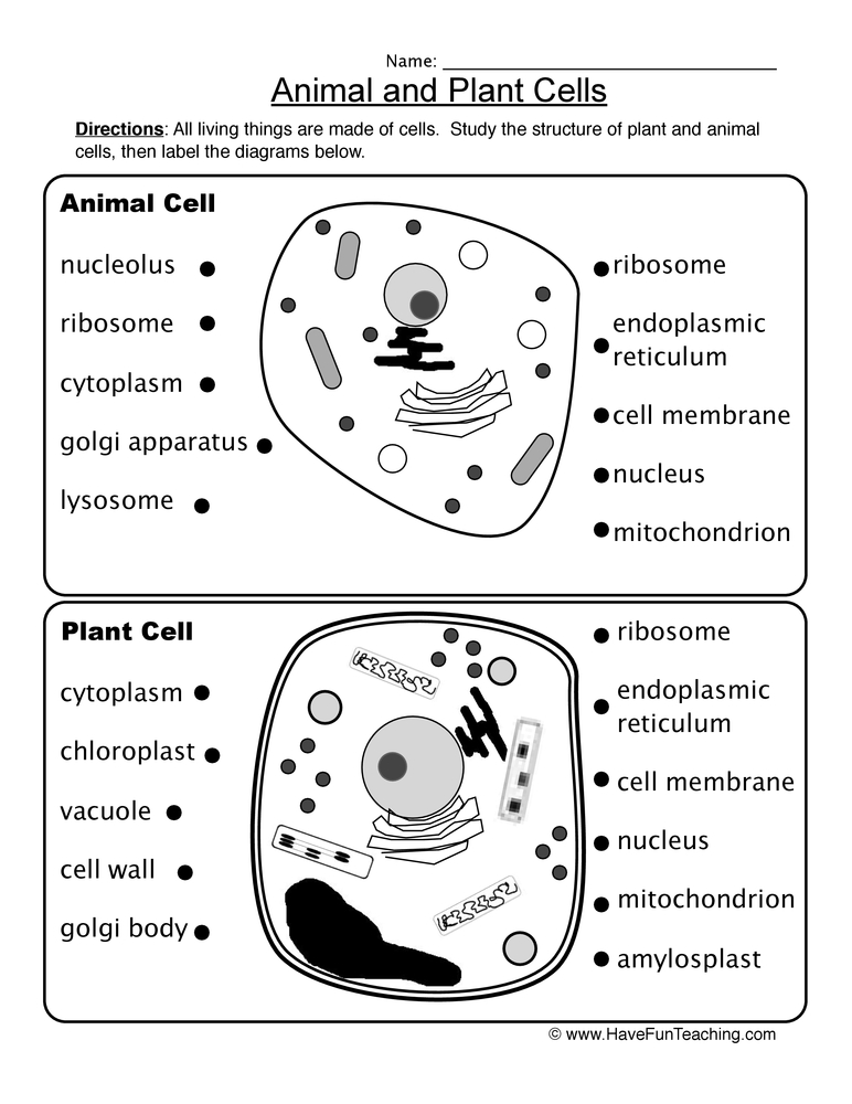 Animal And Plant Cells Worksheet Have Fun Teaching
 Putative Lysosomes Of The Mdck Cell A B And C Show Typical
 Plant And Animal Cells Not Labeled Google Search Animal Cell
 4 Ways To Make An Animal Cell For A Science Project Wikihow
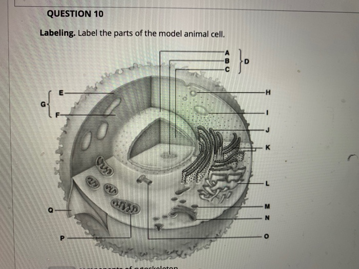 Solved Question 10 Labeling Label The Parts Of The Model
 Rna Granules Hitchhike On Lysosomes For Long Distance Transport
:max_bytes(150000):strip_icc()/animal-cells-vs-plant-cells-373375_final-5b462d7fc9e77c00375014f1.png) Differences Between Plant And Animal Cells
Differences Between Plant And Animal Cells
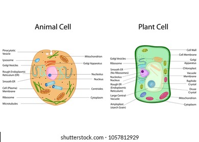 Lysosome Images Stock Photos Vectors Shutterstock


Komentar
Posting Komentar