Labeled Small Intestine Histology Diagram
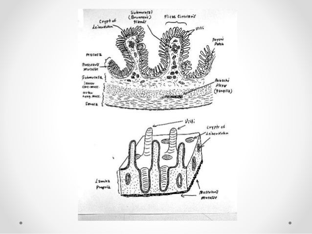
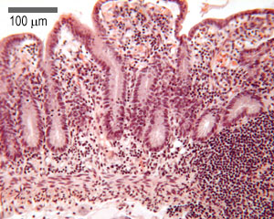
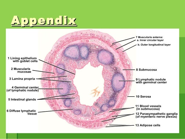
Histological diagram of lateral section tongue large intestine labelled diagram histology of the gastro intestinal tract the mesentery also supplies large intestine with blood from superior and inferior mesenteric arteries histology. The histology of the wall of the small intestine differs somewhat in the duodenum jejunum and ileum but the changes occur gradually from one end of the intestine to the other.
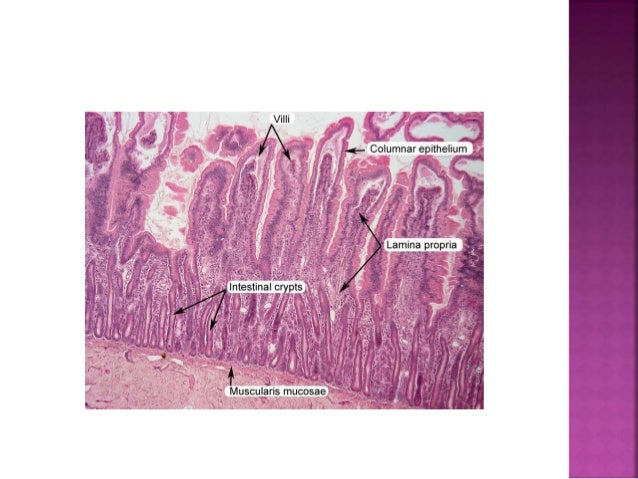
 Small Intestine Histology Labeled Histology Slides Human
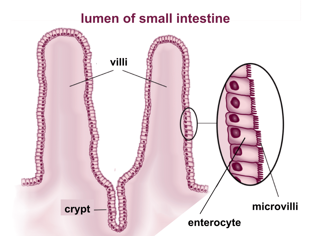
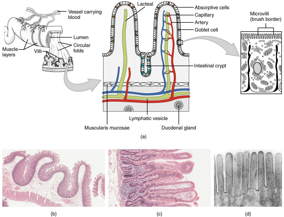
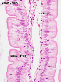
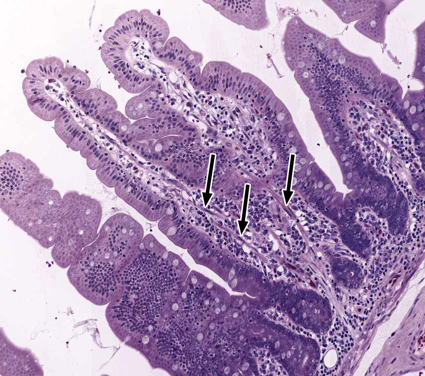
A the absorptive surface of the small intestine is vastly enlarged by the presence of circular folds villi and microvilli.

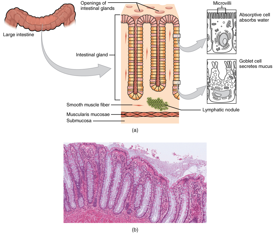
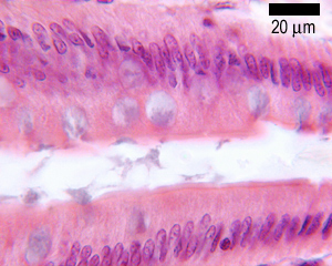
Labeled small intestine histology diagram. In living humans the small intestine alone measures about 6 to 7 meters long. D electron micrograph of the microvilli. The large intestine does not have villi that the small intestine has.
The pancreas and liver also deliver their exocrine secretions into the duodenum. Lymphocytes are small leukocytes white blood cells that are unique to lymphatic tissues. The large intestine also contains lymphocytes that aid in immunity.
The small intestine is made up of the duodenum jejunum and ileum. Musc tube that transmit urine via peristaltic waves leads from kidney is the most posterior structure that emerges from hilus of kidney 25 30 cm long enter bladder at anteromedially superior the anatomy histology and development of the ureter urinary vesicle and urethra. The small intestine is a organ located in the gastrointestinal tract which assists in the digestion and absorption of ingested food.
Anatomically the small bowel can be divided into three parts. Taenia coli are three muscular strips that line the entire large intestine. It extends from the pylorus of the stomach to the iloececal junction where it meets the large intestine.
Together with the esophagus large intestine and the stomach it forms the gastrointestinal tract. Their nerve branches extend throughout the entire length of the small intestine in the form of two plexuses. B micrograph of the circular folds.
Duodenum slide 162 40x pyloro duodenal junct he webscope imagescope slide 161 40x pylorus duodenum pancreas. The small intestine or small bowel is an organ in the gastrointestinal tract where most of the end absorption of nutrients and minerals from food takes place. The small intestine secretes enzymes and has mucous producing glands.
Ureter retroperitoneal general info. From left to right lm x 56 lm x 508 em x 196000. Small intestine innervation diagram the small intestine is innervated by branches of the vagus nerve cn x and thoracic splanchnic nerves.
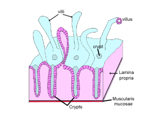
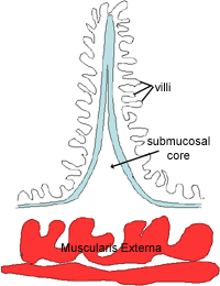
Histology of the small intestine. It is also much more muscular. The duodenum the shortest is where preparation for absorption through small finger like protrusions called villi.
The mucosa is highly folded. The small intestine has three distinct regions the duodenum jejunum and ileum. Large circular folds called plicae circulares shown in the diagram to the right most numerous in the upper part of the small intestine.
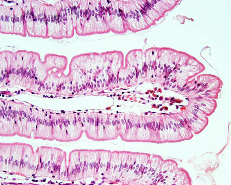
C micrograph of the villi. The duodenum jejunum and ileum. It lies between the stomach and large intestine and receives bile and pancreatic juice through the pancreatic duct to aid in digestion.
After death this length can increase by up to half.
 Small Intestine Histology Labeled Google Search Stomach
 Image Result For Small Intestine Duodenum Histology Labeled
 Small Intestine Structure Histology Secretions
 Small Intestine Histology Human Anatomy And Physiology
Blue Histology Gastrointestinal Tract
 23 5 The Small And Large Intestines Anatomy And Physiology
Blue Histology Gastrointestinal Tract

 Small Intestine Histology Labeled Buscar Con Google Cell
Http Gsm Utmck Edu Surgery Documents Smallintestine Pdf
 File Intestine Histology 002 Jpg Embryology
 Dictionary Normal Duodenum The Human Protein Atlas
 Histology Of The Small Intestine At Magnification 50x 200x And
 Large Intestine Histology Slides Pathology Study Cell Diagram
 Histological Examination Of Rat Small Intestine Taken From The

 Histology Of Duodenum Manage Your Time 1996
Large Intestine Histology Labeled
Blue Histology Gastrointestinal Tract
 85 Best Histology Images Anatomy Physiology Physiology
 Stomach Histology Labeled Histology Slides Physiology Medical
The Small Intestine Boundless Anatomy And Physiology
 Histological Analyses Of Slices From The Small Intestine Of Mice
 Small Intestine Histology Histology Slides Human Anatomy And
 23 5 The Small And Large Intestines Anatomy And Physiology
 Thoc1 Gene Deletion Affects The Histology Of The Small Intestine
 Small Intestine Function Location Parts Diseases Facts
 Histology Of Duodenum Manage Your Time 1996
 Histology Duodenum Small Intestine Histology Duodenum Labels
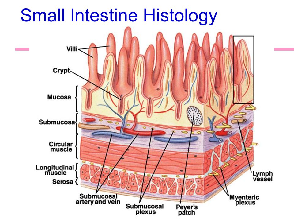 Anatomy Of The Digestive System Ppt Video Online Download
 Layers Of The Small Intestine Youtube
 Histology Digestive Page 1 Frame
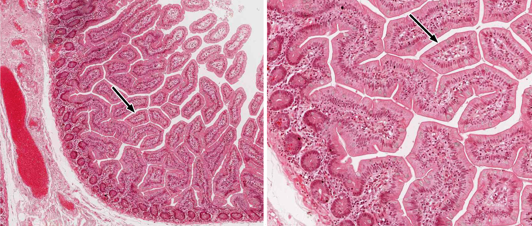 Small And Large Intestine Histology
 Intestine Histology Necrosis Is Observed In Intestinal Microvilli
The Small Intestine Boundless Anatomy And Physiology
 Histology Digestive Page 1 Frame
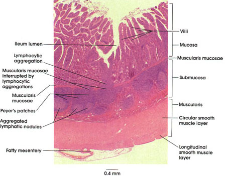 Anatomy Atlases Atlas Of Microscopic Anatomy Section 1 Cells
 Shotgun Histology Small Intestine Ileum Youtube
 Histopathology Of The Small Intestine H E Stained Tissue Sections
:watermark(/images/watermark_5000_10percent.png,0,0,0):watermark(/images/logo_url.png,-10,-10,0):format(jpeg)/images/atlas_overview_image/26/gT2XjjHLHrgNuuhlY1FYKg_structures_of_duodenum_english.jpg) Duodenum Anatomy Histology Composition Functions Kenhub
Duodenum Anatomy Histology Composition Functions Kenhub
Large Intestine Drawing At Getdrawings Free Download
 Histology Digestive Page 1 Frame
 Small Intestine Morphology Of Polgd257a Mice Hematoxylin And
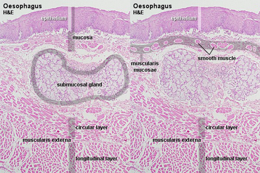 Https Encrypted Tbn0 Gstatic Com Images Q Tbn 3aand9gcr8rqxnxwuvnug0sc9czvftndo1x7vhtyuzyj56f Me P2sakse
:background_color(FFFFFF):format(jpeg)/images/library/7610/OE14P25VJcN0bPWhEvGjg_Intestinal_Villi.png.jpg) Lower Digestive Tract Histology Kenhub
Lower Digestive Tract Histology Kenhub
 45 Best Histology Small Intestine Images In 2020 Histology
 Histology Of Gastrointestinal Tract
 Gastrointestinal Tract Colon Histology Embryology
 A F Representative Histological Sections Of Small Intestine In
Blue Histology Gastrointestinal Tract
 The Small And Large Intestines Anatomy And Physiology Ii
 Stomach Histology Labeled Muscularis Externa Mucosa And
 Histology Of The Distal Ileum A B And Colon C D Of Preterm
 Histology Digestive Page 1 Frame
 Gastrointestinal Tract Intestine Development Embryology
 Small And Large Intestine Histology
Chapter 14 Page 12 Histologyolm 4 0
 Solved 560 Chapter Twenty Five The Digestive System Ceils
 Histology Digestive Page 1 Frame
Gross And Microscopic Anatomy Of The Large Intestine
 Solved 73 Of 113 Histology Of The Small Intestine Duoden
 The Small Intestine Canadian Cancer Society
 Ch23 General Digestive Histology
 Gastrointestinal Tract Wikipedia
 Laminin A5 Influences The Architecture Of The Mouse Small
:watermark(/images/logo_url.png,-10,-10,0):format(jpeg)/images/anatomy_term/duodenum-8/SNgzSimA2NzrRtu5kI8IQ_Duodenum.png) Duodenum Anatomy Histology Composition Functions Kenhub
Duodenum Anatomy Histology Composition Functions Kenhub
Histology Digestion Lab Ileum Peyer S Patch
 Small Intestine Function Location Parts Diseases Facts
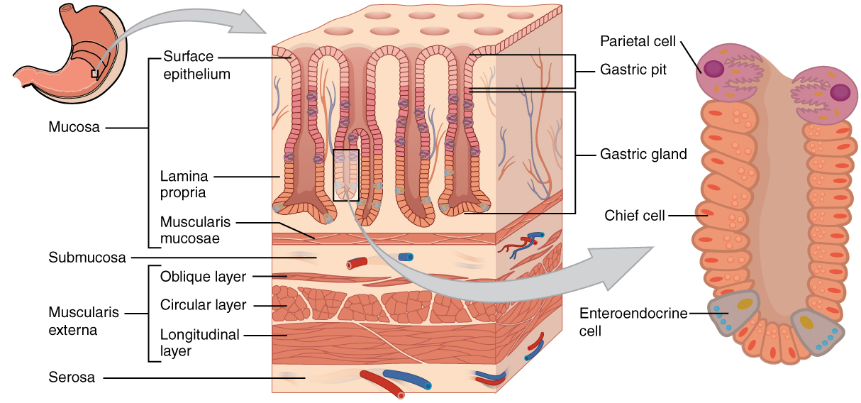 23 4 The Stomach Anatomy And Physiology
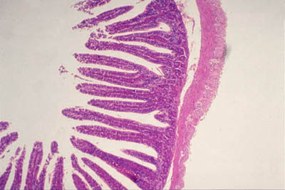 Histology World Key Histology Features
 Small And Large Intestine Histology
Biology W2501 Contemporary Biology Lab Histology
Blue Histology Gastrointestinal Tract
 Histology Digestive Page 1 Frame
 Normal Colon Tissue Video Colon Diseases Khan Academy
:background_color(FFFFFF):format(jpeg)/images/library/12462/structure-of-large-intestine_english.jpg) Colon Anatomy Histology Composition Function Kenhub
Colon Anatomy Histology Composition Function Kenhub
 Histopathology Colon Adenocarcinoma Youtube
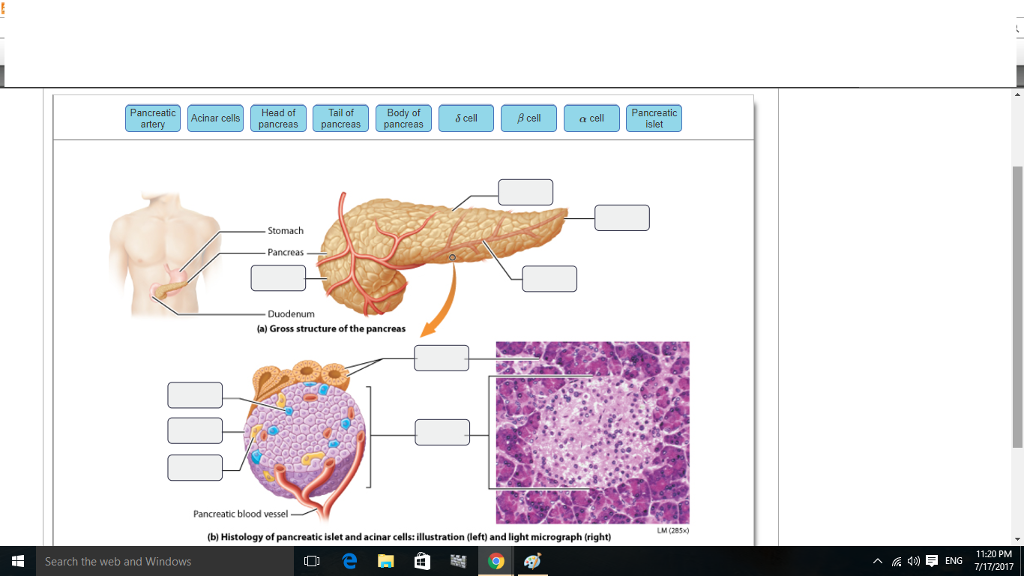 Solved Art Labeling Activity Anatomy And Histology Of Th
 The Small And Large Intestines Anatomy And Physiology Ii
 Ch23 General Digestive Histology
Hepatic Histology Extrahepatic Biliary System
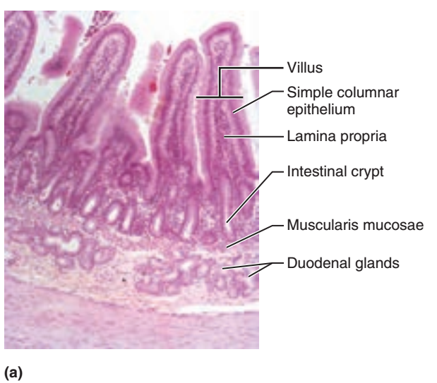 Activity 3 Observing The Histologic Structure Of The Small



Komentar
Posting Komentar