Human Egg Cell Diagram Labeled
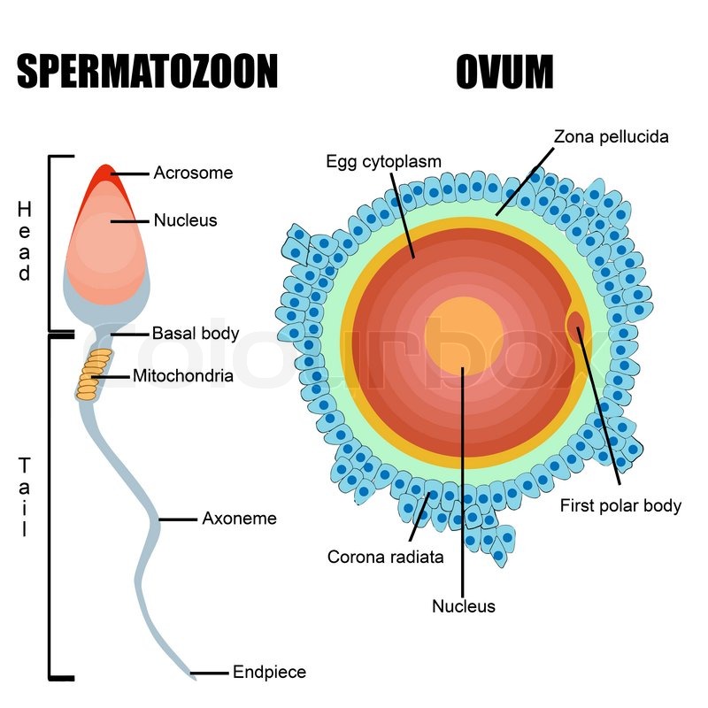
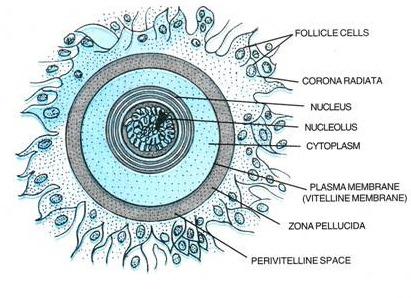
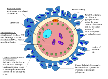
Although diagrams of egg cells commonly include a haploid nucleus no nucleus will form within the egg until after fertilisation has occurred the egg cell is arrested in metaphase ii until it becomes fertilised by a sperm. It contains most of the genetic material in the form of chromosomes.
For example labeled parts of the cactus flower include the stigma the stamen and the anther.

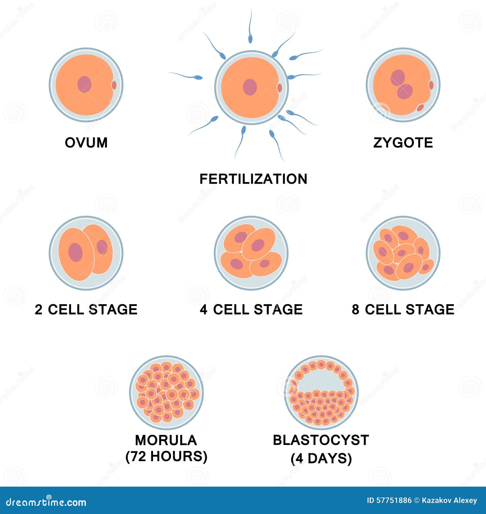
Human egg cell diagram labeled. When egg and sperm fuse a diploid cell the zygote is formed which rapidly grows into a new organism. The nucleus is the heart of the egg cell. In contrast the reproductive cells used by males are referred to as sperm.
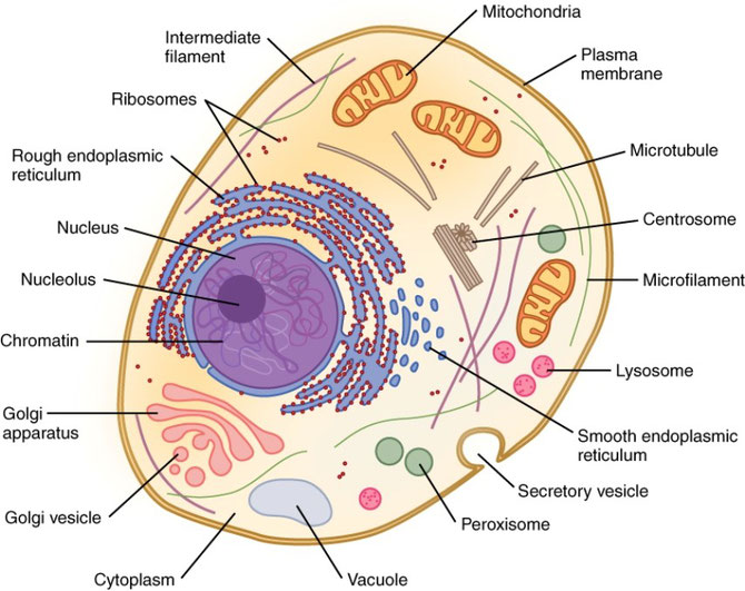
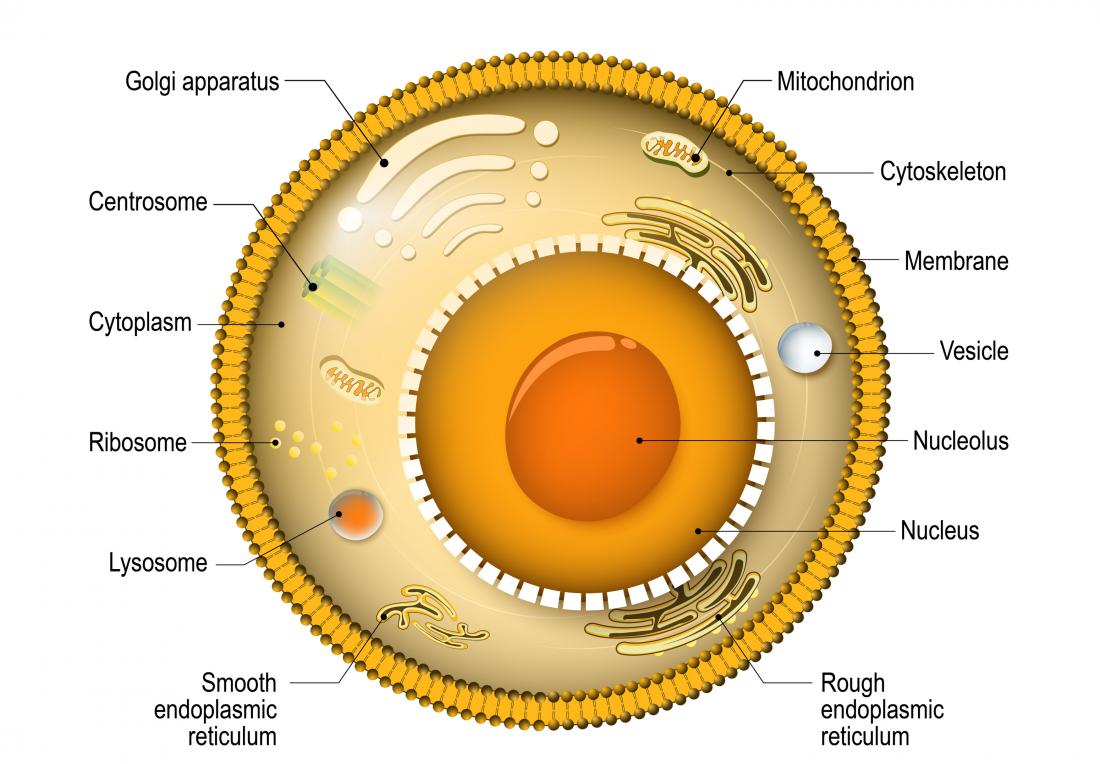
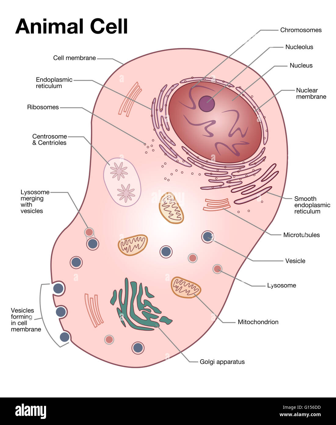
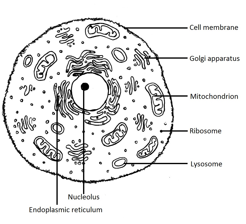
With its spheric shape it consists of a large nucleus which at the same time contains an acrosome. Diagram of the human cell illustrating the different parts of the cell. The cell membrane is the outer coating of the cell and contains the cytoplasm substances within it and the organelle.
This page explains what women donate when they become egg donors. The nucleus contains the genetic information and 23 chromosomes. Human cell properties diagram parts pictures structure last updated on september 7 2018 by surekha the cell is the basic unit of any living organ and it is the organ that replicate on its own determining growth.
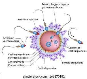
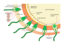
The cell membrane changes after fertilisation by a single sperm so that no more sperm can enter. Egg are larger than any other cell in the human body at about 100 microns or millionths of a meter in diameter about the same as a strand of hair. The haploid nucleus contains the genetic material for fertilisation.
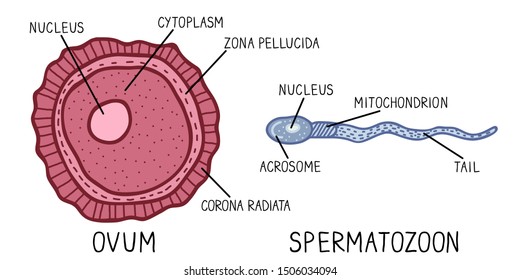
The corona radiata is an external layer of follicular cells which provide support and nourishment to the egg cell. Structure and parts of a sperm cell. Egg cells or ova are the cells utilized by female organisms to reproduce offspring.
The diagram breaks each part of the cactus down to its smallest known level of existence. The egg is one of the biggest cells in the body. Egg cell fact 1.
The egg cell is typically not capable of active movement and it is much larger visible to the naked eye than the motile sperm cells. In mammals a new individual is formed when an egg from the mother and a sperm from the father come together and allow their genetic material to fuse. Within the egg cell are numerous cortical granules which release their contents upon fertilisation to prevent polyspermy.
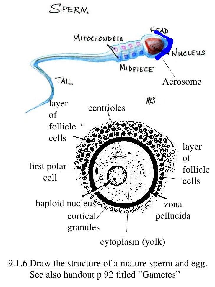
Above you will see a diagram that labels the main parts of the human egg together with an illustration of a real human egg. The egg cell or ovum plural ova is the female reproductive cell in oogamous organisms. That means you could in theory see an egg cell with the naked eye.
It is a double layered membrane composed of proteins and lipids. A labeled part of a cactus is a body part that can be identified on a diagram of the inside and outside of the cactus. This labelled diagram shows the structure of a sperm cell in detail which has the following parts.
The cytoplasm contains nutrients for the growth of the early embryo. Eggs are one of the biggest cells in the body and only a few are made.
 Human Egg Cell Diagram Labeled Cell Diagram Egg Cell Diagram
 Draw A Labeled Diagram Of Human Ovum Released Afte Toppr Com
 Structure Of Egg And Sperm Alila Medical Images
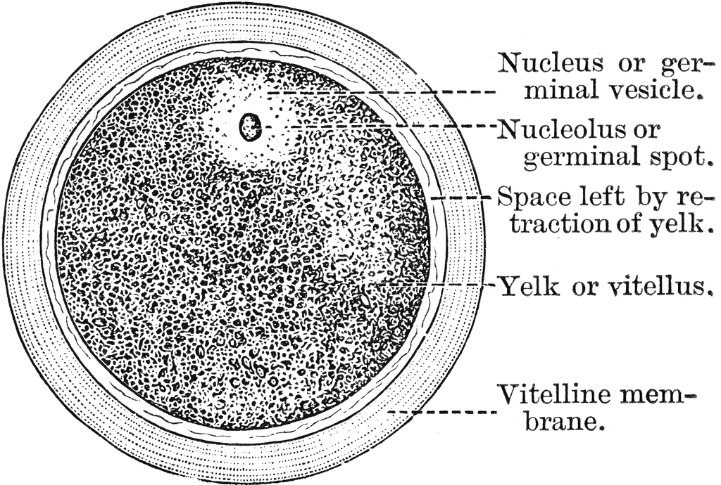 A Diagram Of The Human Ovum Clipart Etc
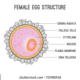 1000 Acrosome Stock Images Photos Vectors Shutterstock
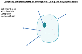

 The Human Egg Cell Explained For Egg Donors Altrui
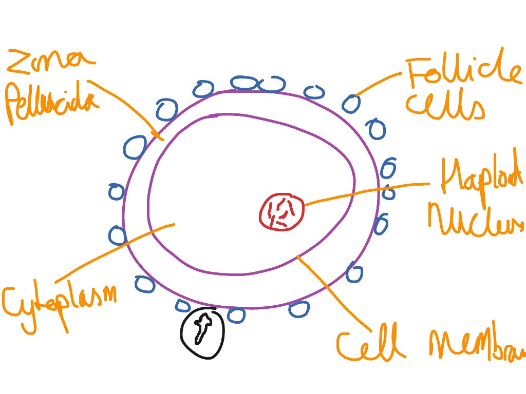 Sperm And Egg Cell Diagram Science Biology Genetics Cells
 Structure Of Human Gametes Egg And Stock Vector Colourbox
 Ovum Images Stock Photos Vectors Shutterstock
 28 Best Egg Cell Human Ovum Images Geek Gifts Learning Tools Ova
Structure Of An Ovum Explained With Diagram
 Animal Cell Parts And Functions Withcarbon
 The Structure And Function Of The Ovum In The Female Reproductive
 Ovum Definition Function Structure Video Lesson Transcript
Embryology The Human Egg Cell And Sperm
 Simple Plant Cell Diagram Labeled Printable Diagram Plant Cell
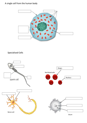
 Ovum Definition Function Structure Video Lesson Transcript
 Pin On This Is When The Magic Happens
 Diagram Showing A Mature Human Spermatozoon With Its Different
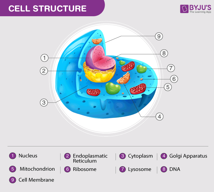 Animal Cell Structure Function Diagram And Types

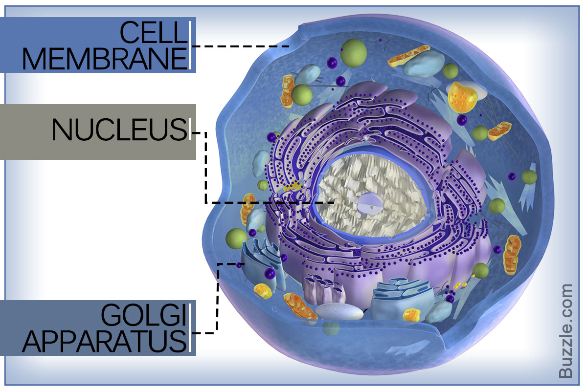 A Labeled Diagram Of The Animal Cell And Its Organelles Biology Wise
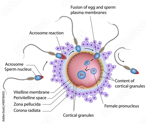 Fertilisation And Polyspermy Block Labeled Buy This Stock
 Structure And Parts Of A Sperm Cell
 Structure Of A Typical Animal Cell Animal And Plant Cell
 Draw The Diagram Of Human Sperm And Label Its Part Toppr Com
 Human Cell Drawing At Paintingvalley Com Explore Collection Of
 Blastocyst Vector Illustration Infographic Diagram Biological
 How Is The Body Of A Sperm Suited For Fertilization Of An Egg
Sexual Reproduction Pass My Exams Easy Exam Revision Notes For
 Human Reproductive System Definition Diagram Facts Britannica
 Stem Cells Vector Illustration Medical Labeled Diagram With
Human Cell Drawing At Getdrawings Free Download
 Structure Of The Egg Incubation And Embryology University Of
Female Reproductive System Archives Medical Information Illustrated
 1000 Acrosome Stock Images Photos Vectors Shutterstock
 The Cell Types Functions And Organelles
 Development Of The Human Embryo Stock Vector Illustration Of
Embryology The Human Egg Cell And Sperm
 Baisc Building Block A Cell Atchaya Paathiram
 Ib Biology Draw Assessment Statements

 Human Ovum Structure The Cell Theory Of Spirituality Science Of
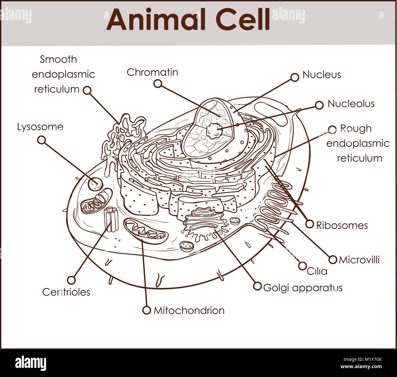 Animal Cell Diagram Stock Photos Animal Cell Diagram Stock
 Cytoskeleton Definition Structure Function With Diagram
3 3 Eukaryotic Cells Concepts Of Biology 1st Canadian Edition

Diagram Onion Cell Diagram Parts Labeled Full Version Hd Quality
 B5 Mitosis Meiosis Exam Practice And Mark Scheme
 Stem Cell Basics A Closer Look At Stem Cells
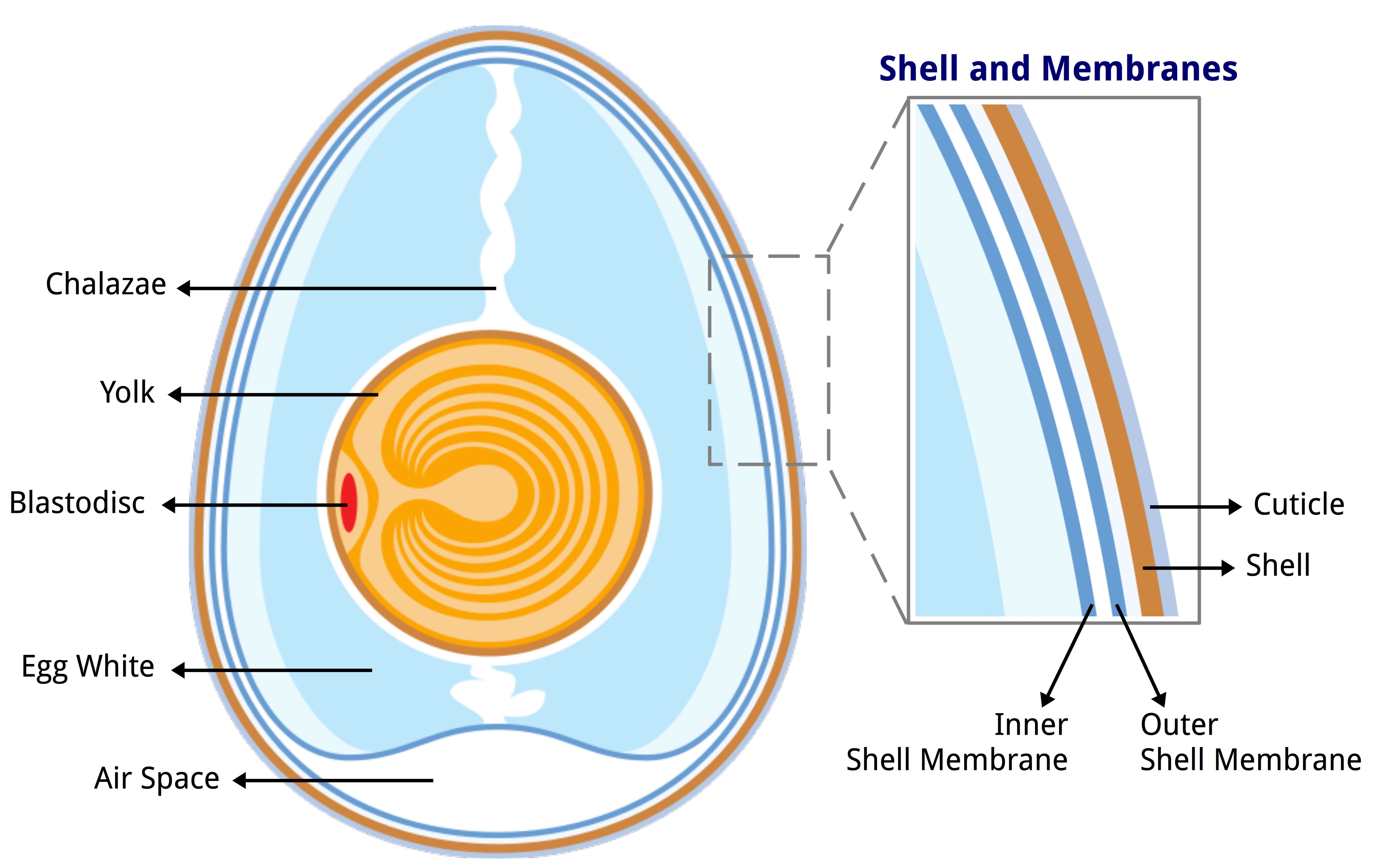 File Anatomy Of An Egg Labeled Jpg Wikimedia Commons
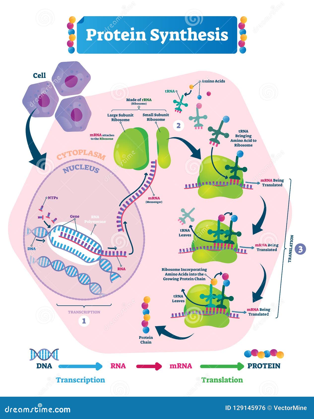 Protein Synthesis Vector Illustration Transcription And
 Anatomy And Physiology Of Animals The Cell Wikibooks Open Books
 Cytoskeleton Definition Structure Function With Diagram
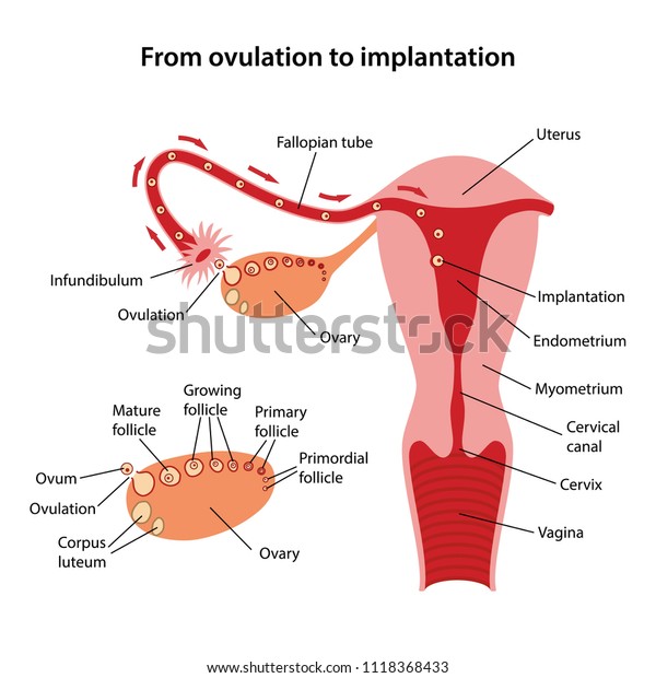 Diagram Process Ovulation Implantation Human Embryo Royalty Free
 Appliance Science The Biology Of The Chicken Egg Cnet
 Animal Cell Diagram Stock Photos Animal Cell Diagram Stock
 The Histology Guide Female Reproductive System
 Animal Cells Do Not Have Cell Walls Biological Science Picture
 Ovary Animal And Human Britannica
Embryology The Human Egg Cell And Sperm
 A Labeled Diagram That Explains The Function Of Nucleolus

 Jorans Lille Skrivebord Health The Cell
Cell Division Mitosis And Meiosis Biology 1511 Biological
Animal Cell Diagram Labeled For Kids Cliparts Co
 The Cell Cycle Mitosis And Meiosis University Of Leicester
 Flowering Plant Reproduction Ii
 Early Human Development Alila Medical Images
 Idea For Green Schools Sub Brand Cell Diagram
 Diagram Of Human Egg Wiring Diagram Blog
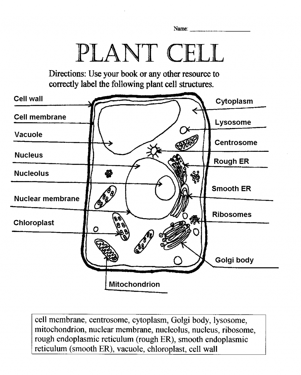 5 Label Plant Cell Worksheet Biological Science Picture
Diagram Labeled Diagram Egg Cell
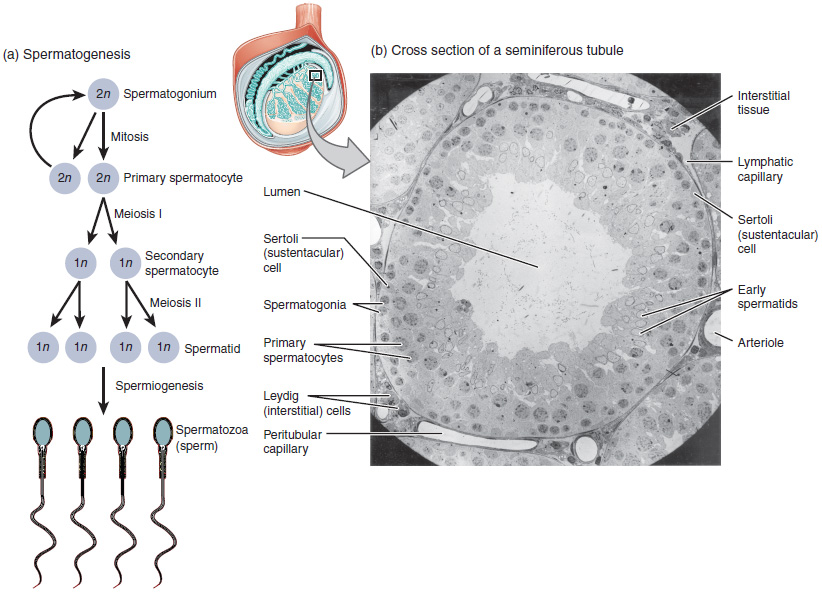 27 1 Anatomy And Physiology Of The Male Reproductive System
 Fertilization Mechanisms In Flowering Plants Sciencedirect
Https Www Studocu Com En Us Document Rochester Community Technical College Elements Of Biology Assignments Mc Graw Hill Education Connect Biology Chapter 89 Hw 2623624 View
 Stem Cell Basics A Closer Look At Stem Cells
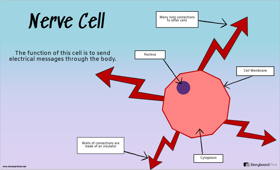 Specialized Cells Activities Cell Structure And Function





Komentar
Posting Komentar